Elucidation of the mechanism of action of EO*
Step1: Uptake from the intestinal tract Absorbed into the body via Peyer’s patches
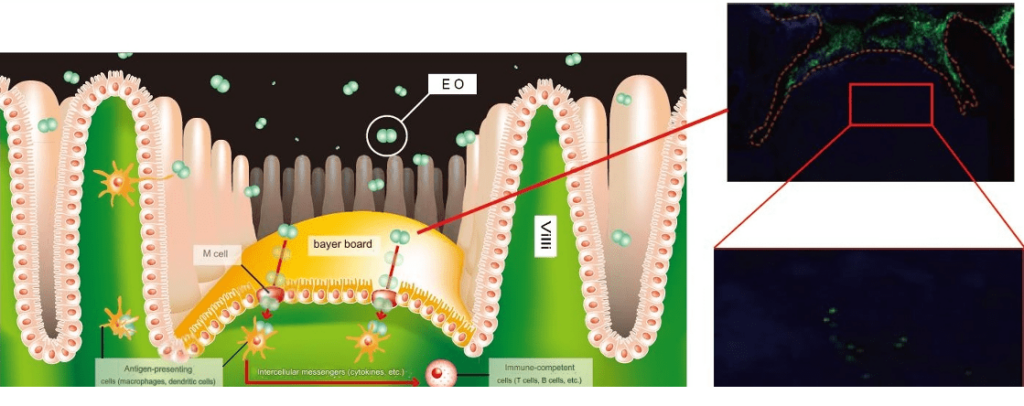
Step2: Phagocytosis by antigen-presenting cells (macrophages and dendritic cells)
Mannose present in the cell wall is recognized and phagocytosed by antigen-presenting cells.
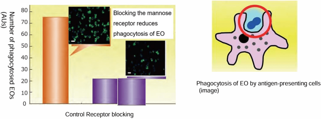
Step 3: Cytokine production by antigen-presenting cells
The phagocytosed EO RNA changes the amounts of cytokines produced by antigen-presenting cells.
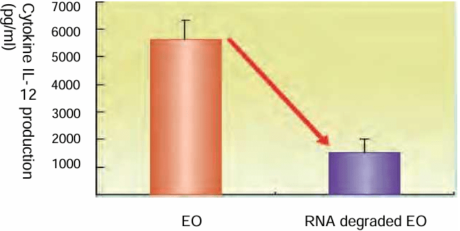
IL-12 production by antigen presenting cells that phagocytosed RNA-degrading EO was significantly reduced. From this, it is speculated that antigen presenting cells are activated by the RNA present in phagocytosed EO, which may be effective against various immune diseases.
*EO = ingredient of Eating Oxygen
Activation of innate and adaptive immunity
[Background]
Previous research has revealed that administration of EO has a suppressive effect against infection with
drug-resistant bacteria. An increase in drug-resistant bacteria-specific IgG in the blood (activation of
adaptive immunity) was confirmed, but because the biological defense effect was confirmed earlier than
the activation of adaptive immunity, innate immunity was also evaluated.
[Method]
30 newly pregnant broiler chicks (1 day old) were divided into two groups and raised on normal feed and
feed containing 0.05% (w/w) EO. Seven chicks were euthanized at 5 days of age and the remaining
eight chicks at 15 days of age, and the gene expression level of 6-defensin2 in the Fabricius sac1 and
the IgA concentration in the cecal contents were measured.
*1 The Fabricius is an immune organ unique to birds, located near the cloaca, and its main role is to
differentiate precursor cells into B cells.
*2 Defensin is a general term for antibacterial proteins (peptides) secreted by cells and is known as one
of the body’s defense mechanisms against bacterial infections.
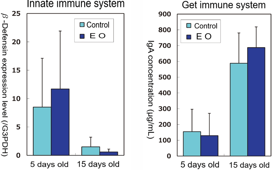
[Results and Discussion]
Immediately after hatching, that is, at the stage when the cells first encounter various foreign
substances, innate immunity is activated, and it was observed that adaptive immunity is gradually
activated. Since the gene expression level of 6-defensin in the Fabricius and the IgA concentration
in the cecum tended to increase with administration of EO, the effect of EO in suppressing infection
by drug-resistant bacteria can be explained by the activation of innate and adaptive immunity at an
effective timing.
Promotes recovery from influenza
[Method]
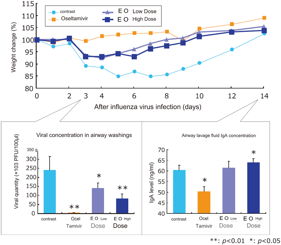
6-week-old BALB/d mice were orally administered 1 mg or 5 mg of EO and oseltamivir from 7 days before
to 7 days after virus infection, and the test period was up to 14 days after virus infection. The virus used
was the A/NWS/33 strain (H1N! subtype). Three days after infection, airway lavage fluid and lungs were
collected, and the amount of virus was measured. 14 days after infection, neutralizing antibody titers in
serum and airway lavage fluid, as well as the amount of virus-specific secretory IgA in airway lavage fluid
and intestine were measured. In addition, weight changes and symptoms were observed throughout the
entire period.
[Results and Discussion]
In the control group, weight loss occurred due to influenza infection, but in the EO-administered group,
weight loss was suppressed and the weight returned to pre-infection levels after 10 days. In addition,
compared to the control group, the EO-administered group showed a significant decrease in the amount of virus in airway lavage fluid in an EO dose-dependent manner, and a significant increase in the amount of influenza virus-specific IgA.
Oral intake of EO is expected to have an effect of reducing viral infections through the body’s defense
mechanism.
Anti-inflammatory effect in mice (contact dermatitis model)
[Method]
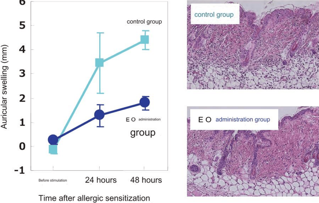
Four-week-old BALB/c mice were divided into an EO intake group (equivalent to 10 mg/kg/day, freely
available) and a normal diet intake group, and contact hypersensitivity was induced and compared. At 16
weeks of age, each group was sensitized with 50 pL of 0.5% dinitrofluorobenzene (DNFB) solution (with a
spatula) to induce contact hypersensitivity, and 5 days after sensitization, 20% of 0.2% DNFB solution was
applied to the ear to induce a contact hypersensitivity reaction. Swelling of the ear was measured with a
dial thickness gauge 24 and 48 hours after hypersensitivity induction.
[Results and Discussion]
Ear swelling was reduced by half in mice fed sterilized lactic acid bacteria EO compared to mice fed
normal feed. Histological staining of the ears revealed that infiltration of inflammatory cells was significantly suppressed in the EO-treated group. It was suggested that the continuous intake of sterilized lactic acid bacteria EO controls allergic reactions that cause inflammation.
Allergy suppression effect (mouse asthma induction model)
[Method]
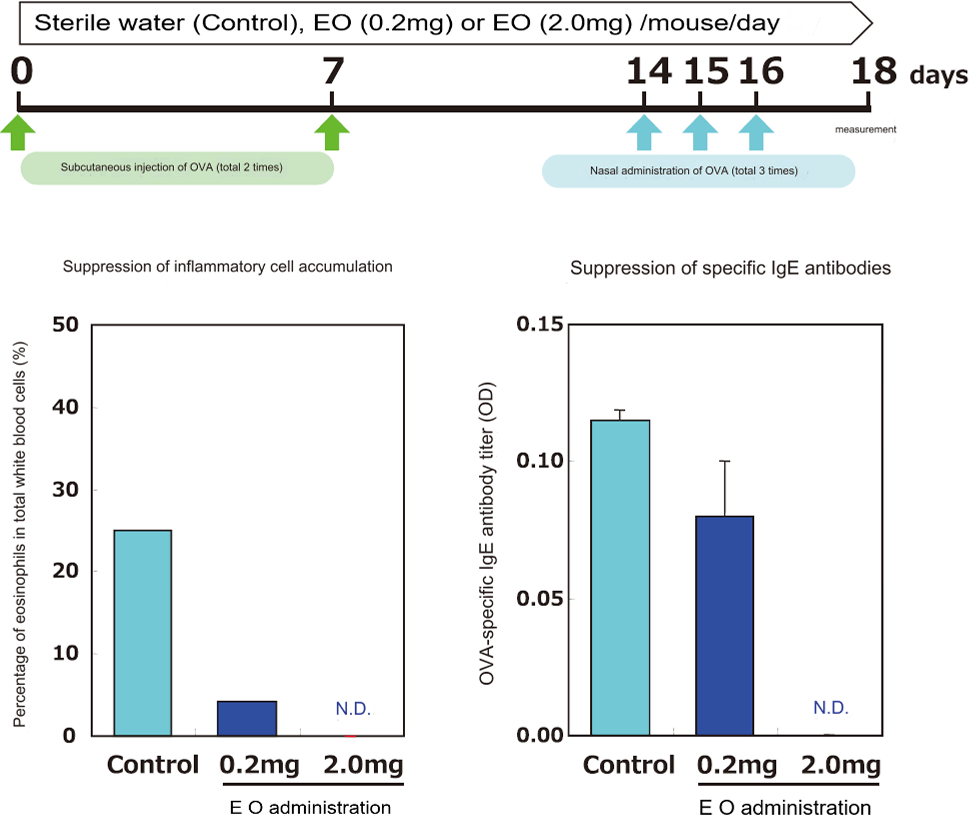
7-week-old BALB/C female mice were sensitized by subcutaneous injection of OVA (antigen) together with an adjuvant, and then OVA was administered intranasally to create an asthma-inducing model. The white blood cell and eosinophil counts in the alveolar lavage fluid, and OVA-specific IgE in the serum were
measured. EO was administered daily throughout the test period. (n = 5)
*OVA: ovalbumin (the substance that causes egg allergies)
[Results and Discussion]
In the control group, accumulation of alveolar eosinophils and an increase in serum antigen-specific IgE
antibody titers, both of which are indicators of allergic symptoms, were observed, but administration of EO caused a dose-dependent decrease in the number of eosinophils and antigen-specific IgE antibody titers, and in the high-concentration administration group, these were suppressed to normal levels.
Ingestion of sterilized lactic acid bacteria EO significantly suppressed the increase in antigen-specific IgE
antibody titers, which are the trigger for allergic reactions, and the accumulation of eosinophils, which
promotes inflammatory reactions, strongly suggesting that EO reduces type I allergic reactions (hay fever,
atopic dermatitis, asthma, etc.).
Improves atopic dermatitis (human clinical trial)
[Method]
150 patients with inflammatory skin diseases were given 400mg of EO per day for more than 2 months,
and continued to take it for diseases (atopic dermatitis) that doctors judged to be effective. The
improvement effect was examined using the changes in blood total IgE levels and antigen-specific IgE
levels (IgE RAST) before and after administration of EO as indicators, and the improvement effect was
evaluated overall by scoring the objective improvement effect by the doctor and the subjective
improvement effect by the patient.
[Results and Discussion]
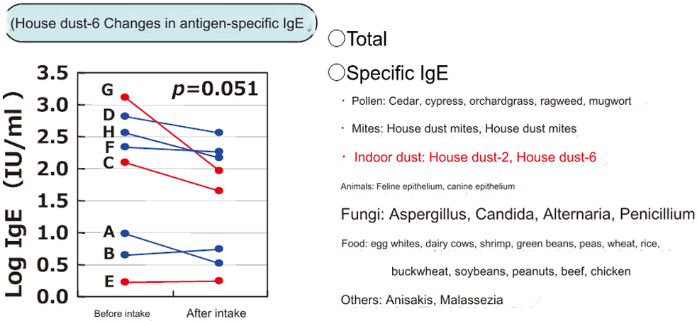
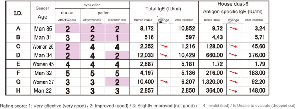
Among the patients with atopic dermatitis, continuous data could be obtained from eight cases. Ingestion
of sterilized lactic acid bacteria EO showed a tendency to suppress the production of specific IgE
antibodies to six house dust antigens, which are likely to be a cause of atopic dermatitis. In addition,
efficacy evaluations by doctors and satisfaction ratings from the patients themselves were obtained,
suggesting an effect on improving the quality of life for patients with atopic dermatitis.
No change was observed in total IgE levels before and after ingestion. On the other hand, a tendency
was observed for antigen-specific IgE levels to decrease after ingestion of EO.
Improves hay fever (human clinical trial)
[Method]
150 patients with inflammatory skin diseases were asked to take 400mg of EO per day for more than two
months, and continued to take it for diseases (hay fever) that the doctor judged to be effective. The
improvement effect was examined using the fluctuation of the total IgE level and antigen-specific IgE
level (IgE RAST) in the blood before and after EO administration as an indicator, and the improvement
effect was scored objectively by the doctor and subjectively by the patient, and the blood was evaluated.
[Results and Discussion]
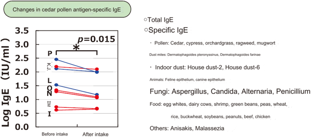
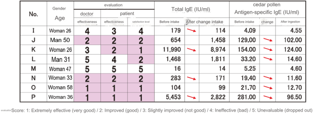
Among the hay fever patients, continuous data could be obtained from eight cases. Ingestion of sterilized
lactic acid bacteria EO significantly suppressed the production of cedar-specific IgE antibodies, which are
a cause of hay fever. In addition, efficacy evaluations by doctors and patient satisfaction were obtained,
suggesting an effect on improving the quality of life for hay fever.
No change was observed in total IgE levels before and after ingestion. On the other hand, a tendency for
antigen-specific IgE levels to decrease after EO ingestion was observed.
Antitumor effects
[Method]
EO was orally administered to 7-week-old BALB/c female mice at a concentration of 0.01mg/kg
or 1mg/kg of mouse body weight every day during the experiment. One week after oral
administration, fibrosarcoma cells, Meth A, were inoculated subcutaneously on the back at a
concentration of 2.0×10⁶/mouse. The survival rate after tumor inoculation was observed.
[Results and Discussion]
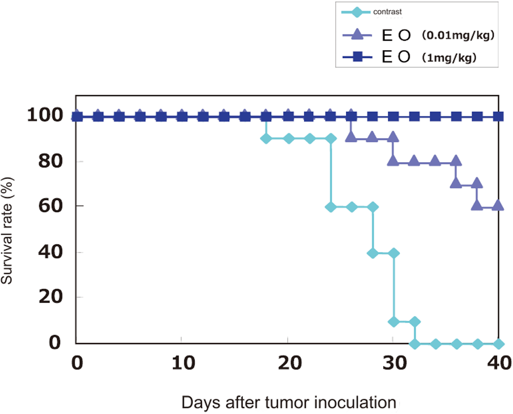
In this study, mice administered saline died between the 18th and 32nd days, but in the group
administered high doses of sterilized lactic acid bacteria EO, not a single mouse died even up to the
40th day of the study period. Furthermore, a higher survival rate was observed compared to the
control group even at a dose 1/100th that of the control group, confirming dose-dependence of the
antitumor effect.
Prevention of intestinal cancer
[Method]
Five-week-old male and female Min mice (a model of human familial adenomatous polyposis) were fed a
diet containing 100 ppm EO for eight weeks, and the carcinogenesis-suppressing effect was examined by
measuring the number and size of intestinal polyps. The control group was fed additive-free AIN-76.
[Results and observations]
EO administration tended to reduce the number of oocyte polyps in both males and females, and a
significant reduction (RV0.05) was observed in the proximal small intestine of male mice.
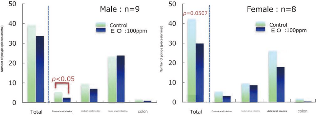
Regarding size and number of intestinal polyps, a tendency for EO administration to reduce the number of small polyps, particularly those less than LO mm, was observed, and a significant (pv0•05) decrease was
observed in the number of polyps less than 0.5 mm and those 0.5 to 1.0 mm in size in females.
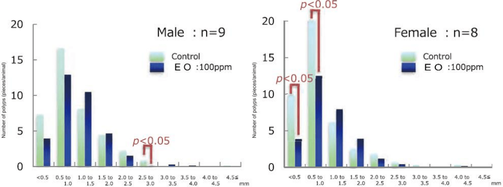
Furthermore, confirmation of gene expression levels of cell proliferation-related factors in intestinal polyps
revealed an inhibitory effect on the activation of the g-catenin signal pathway, which is involved in intestinal
carcinogenesis. Sensitization tests on human colon cancer cell line HCT-116 also suggested that EO
ingredients may act directly. (All data not shown).
The results of this study suggest that EO intake may have an inhibitory effect on intestinal carcinogenesis, and that the mechanism may be different from that of probiotics.
Colitis suppression effect
[Method]
5-week-old C57BL/6NlHfiT live mice were orally administered EO (Low: 10mg/kgBW, High:
100mg/kgBW) and induced colitis by administering 2.5% DSS in drinking water for 7 days from the
14th day of administration.
Mice were dissected on the 18th day of administration, and pathological observations of colon tissue
and gene expression analysis of inflammatory cytokines were performed. Group comparisons were
made between the DW (distilled water) administration group (n = 5), the DSS administration group (n =
5), the EO low group (n = 5), and the EO high group (n = 5).
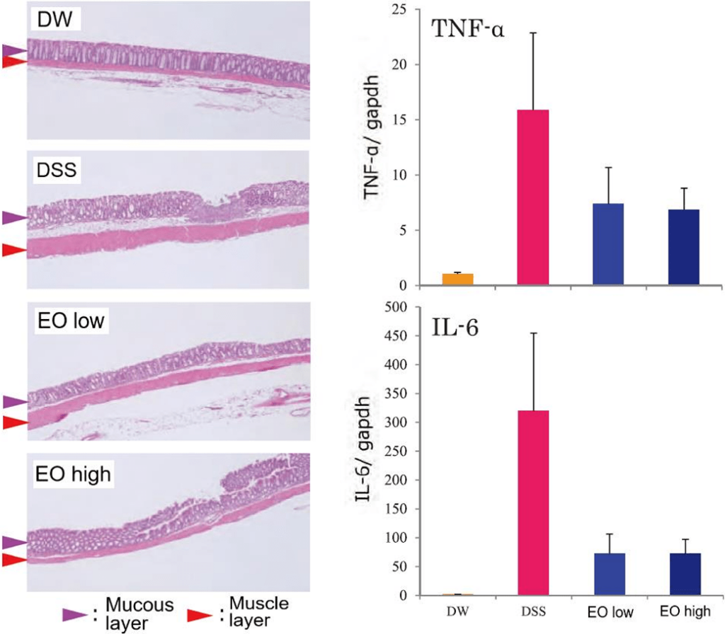
[Results]
Pathological observations showed thickening of the muscular layer and the submucosal layer between
the mucosal layer and the muscular layer in the DSS-administered group, but EO administration tended
to suppress each of these thickenings. In addition, the expression of inflammatory cytokines also tended
to be suppressed in the EO-administered group.
Therefore, EO intake is expected to have an inhibitory effect on colitis.
